Written by Fabio Procoli, Specialist in Small Animal Medicine
What is protein losing enteropathy (PLE)?

PLE of dogs is a syndrome by excessive loss of serum albumin from the intestinal mucosa and wall. The pathogenesis of PLE is complex with several potential causes playing a role such as increased intestinal vascular permeability induced by inflammation; loss of integrity of the intestinal mucosa secondary to ulcers or erosions; dilatation and/or obstruction of intestinal lacteals (lymphatics vessels) secondary to inflammatory or neoplastic processes. Each or a combination of the above mechanisms will result in the intestinal loss of blood proteins, most commonly albumin, which when severe can lead to low serum protein level (hypoalbuminemia).
Based on the available scientific literature we know that in roughly 65% of cases of canine PLE the underlying cause is a chronic inflammatory enteropathy (also known as IBD, which stands for inflammatory bowel disease) and in around 50% of these cases there is also a component of intestinal lacteals disease (lymphangiectasia). Less common causes include endocrine disorders (such as Addison’s disease), neoplastic disease (such as low grade lymphoma) or fungal diseases (reported only in some southern states of North America or in countries with subtropical climate).
Are certain dog breeds more predisposed to PLE than other?
Several studies have shown an association for PLE with certain canine breeds. Historically the most commonly reported breeds include the Yorkshire Terrier, Rottweiler, Maltese and German Shepherd. However, more recent evidence suggests that also Cavalier King Charles together with other breeds (such as Pug) might be at higher risk of developing PLE.
Which symptoms do dogs with PLE show?
Clinical signs in PLE can vary with regards to type and severity whilst some dogs might show no clinical whatsoever at the time of diagnosis. When present the most common clinical signs include:
- Lethargy
- Loss of appetite
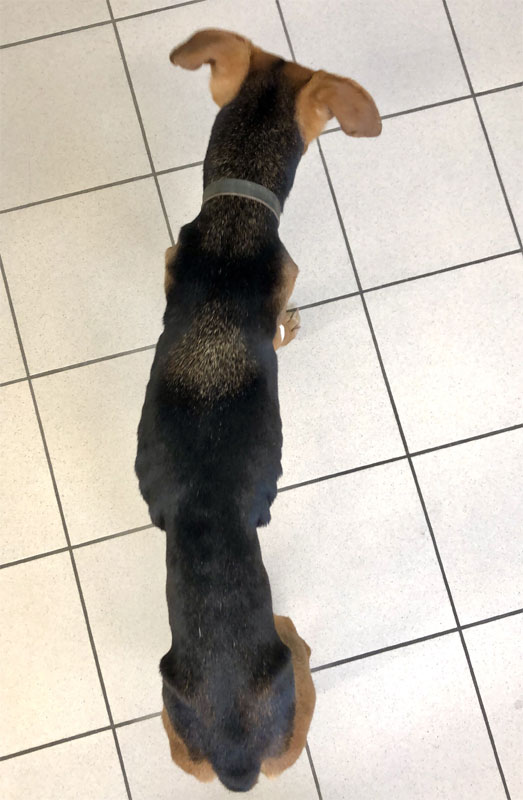
- Loss of body condition or muscle mass (Figure 1)
- Vomiting
- Diarrhoea
- Abdominal pain
- Peripheral oedema
- Abdominal, and less commonly, thoracic free fluid accumulation
How can I know if my dog has PLE?
PLE should be suspected if a dog, especially if belonging to a predisposed breed, shows compatible clinical signs and there is evidence of intestinal protein loss with/or without hypoalbuminemia. In order to further suspect PLE, other causes for the clinical signs and hypoalbuminemia will have to be excluded by means of abdominal imaging (to exclude intestinal masses or neoplasia), endocrine testing (to rule out Addison’s disease) and urinalysis (to rule out protein losing renal disease).

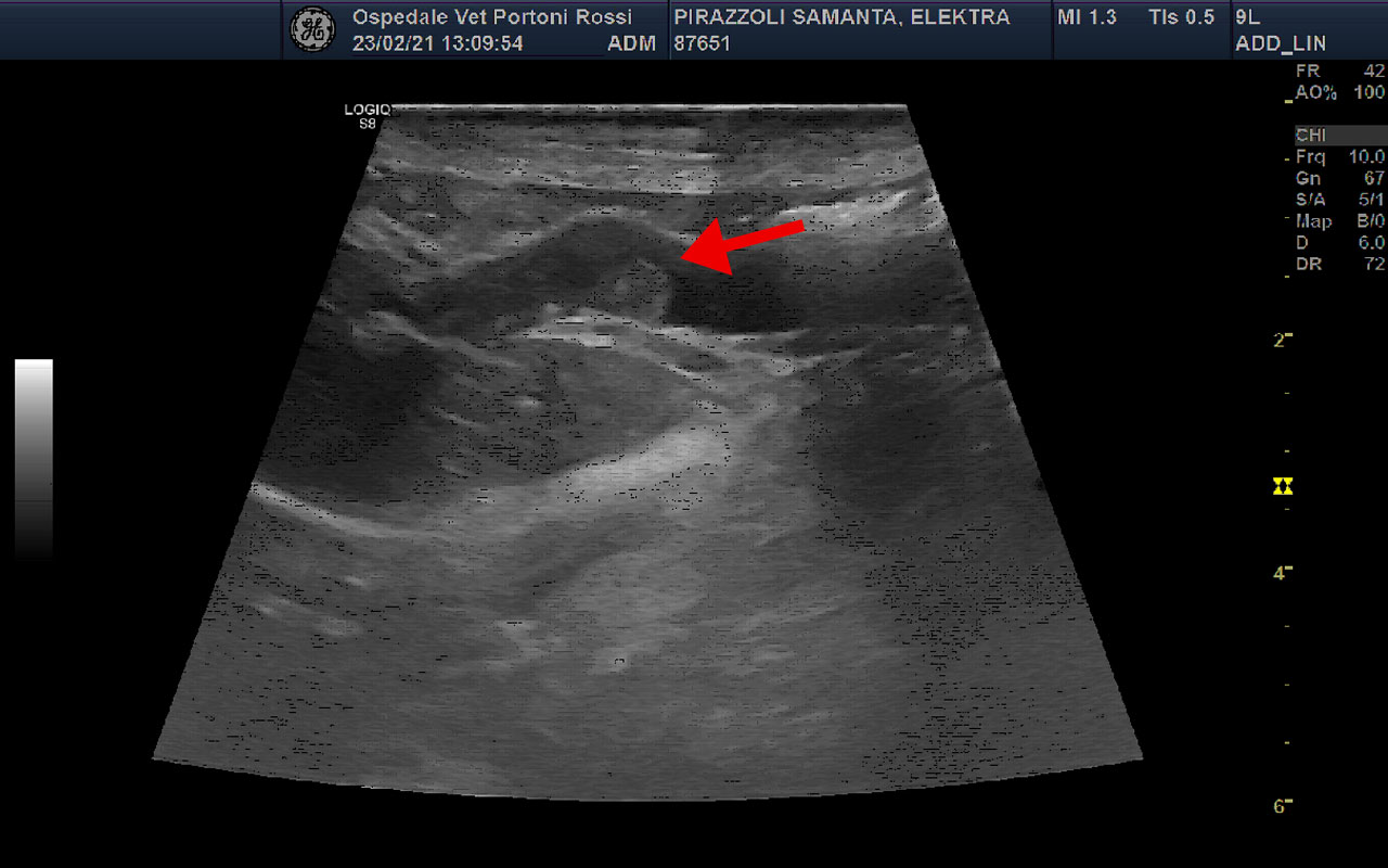
Typical findings suggestive of PLE on abdominal ultrasound include thickening of the small intestinal wall, presence of hyperechoic striations within the intestinal mucosa (Figure 2), mesenteric lymph nodes enlargement, hyperechogenicity of the abdominal lining (peritoneum) and presence of abdominal free fluid (Figure 3).
How can we confirm the diagnosis of PLE and understand its cause?
Histopathological examination of small intestinal biopsies (most commonly collected via endoscopy and less commonly via surgery) is necessary in order to confirm a diagnosis of PLE, to understand its cause and to define a more specific treatment plan. Histopathological abnormalities typical of PLE include mucosal inflammatory infiltrates, injury and/or atrophy of intestinal villi, lacteal dilatation, and distension and/or abscess of the intestinal crypts.
How is PLE treated?
Treatment of PLE will depend on the underlying diagnosis and the findings of intestinal histopathology. Treatment protocols might include dietary fat restriction (by means of commercial low fat diets or homemade ultra-low fat diets, the latter with less than 6% of fat content). If indicated by the findings of histopathology, medical treatment with glucocorticoids (steroids) might be considered. Additional treatments such as anti-platelet and anticoagulant therapy, prebiotics, probiotics, vitamin B12 supplementation or second line immunomodulatory drugs may be considered on a case-by-case basis.
Are there any complications associated to PLE?
Major complications secondary to PLE include pulmonary and systemic thromboembolism (which could be potentially fatal), accumulation of free abdominal and thoracic fluid and loss of body weight and muscle mass.
What’s the prognosis with canine PLE?
The prognosis of canine PLE will depend on the stage and severity of the disease at the time of diagnosis and above all on the underlying cause. Generally speaking, positive response to dietary management and lack of severe clinical signs are good prognostic indicators. On the contrary, lack of response to dietary management, high clinical severity scores or the presence of an underlying neoplasm are negative prognostic indicators.